
One platform
Today, particles below 200 nanometres can only be imaged in specialised laboratories: in a vacuum, after elaborate preparation, usually for a single question. That is expensive: even a benchtop instrument costs 80,000 to 250,000 dollars, a field-emission research instrument half a million euros and more – plus a laboratory, maintenance and specialists to operate it. This is why electron microscopes are mostly found in research institutions and large companies.
Our microscope measures in air and analyses the results itself. Our target price is in the low five figures in euros, a fraction of today’s instruments: compact, without sample preparation and without a dedicated laboratory, operated by an AI instead of specialist staff. That way it can stand where the questions arise – in production, at the measuring station, in the clinic. The applications differ mainly in software, particle material and regulatory effort – which is why we start where no approval is needed.
Industry
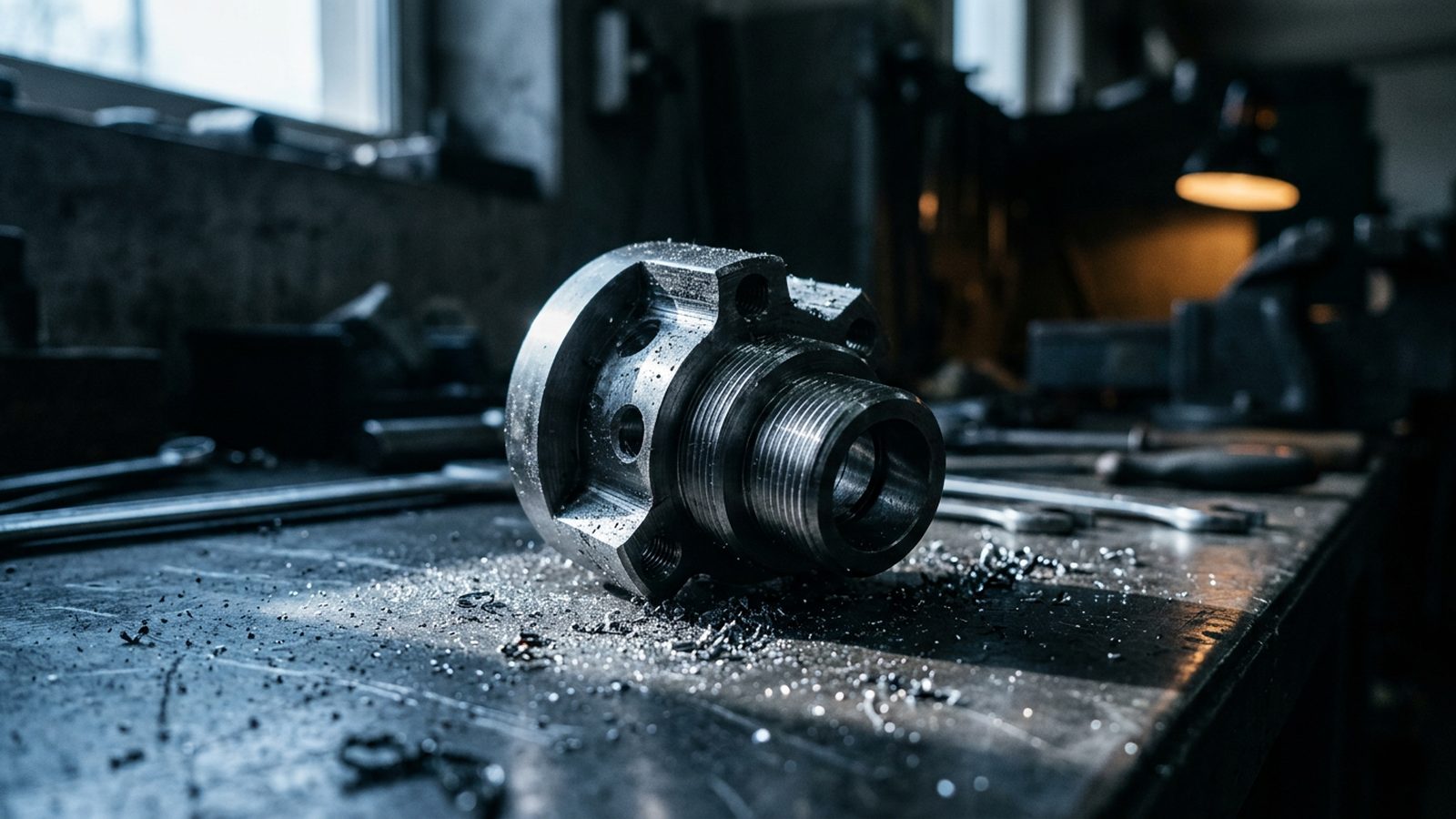
Particles in manufacturing: abrasion from metals and brakes, contamination in semiconductor and battery processes. Seeing every particle individually reveals, from its shape and material, where it comes from – so that the source can be eliminated.
Industrial applications require no regulatory approval. That is why they come first.
Ultrafine dust in the air
Ultrafine dust from traffic, combustion and industry penetrates deep into the lungs and bloodstream. Common instruments count particles or determine their mass – they do not see which particles they are. An imaging measurement shows particles individually rather than averages and adds the question of their origin to today’s methods.

Nanoplastics in water
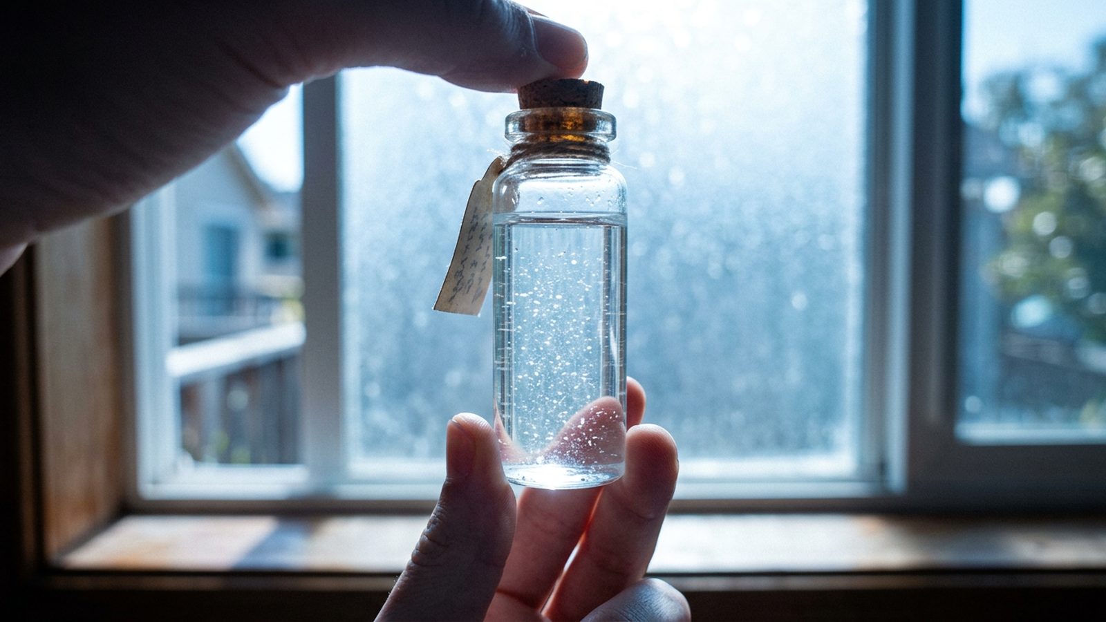
Plastic breaks down into ever smaller particles. Nanoplastics enter the body through drinking water, food and air. Measuring their concentration is laborious with today’s analytical methods. Because our sample can also remain in liquid, the aim is to image plastic particles individually and tell them apart from other particles.
Medicine, in the medium term
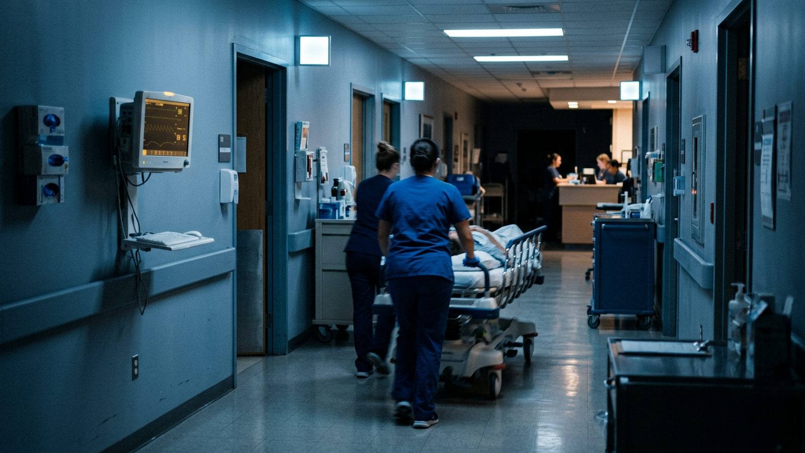
In a severe infection such as sepsis, every hour counts. The usual diagnosis via cultures often takes several days; until then, broad-spectrum antibiotics are frequently given, which promote the development of resistant pathogens. Worldwide, around five million deaths each year are associated with antibiotic-resistant bacteria (estimate for 2019).
Bacteria themselves are large enough for the light microscope. There, however, they only appear as rods or spheres of about one micrometre; after staining they can be roughly sorted into groups, but which species it is usually only becomes clear after growing them in culture. The electron microscope shows shape and surface in detail. How far pathogens can be told apart by these features is something we want to investigate with the prototype.
Our medium-term goal: recognising pathogens directly in the sample. This is a goal that requires regulatory approval, not a current offering.
Research
For universities and laboratories, a compact electron microscope that measures in air and analyses the results itself is a tool for series of investigations that have so far been tied to large instruments and elaborate sample preparation.
For investors
- Approach
- Compact scanning electron microscope that measures in air through a window; our own hardware right down to the beam; a physical model of the instrument
- Cost
- Today's scanning electron microscopes cost 80,000 to 250,000 dollars as benchtop instruments and half a million euros and more as research instruments, plus laboratory, maintenance and specialist staff (R&D World, March 2026). Target price for our instrument: in the low five figures in euros, without a laboratory or specialist staff
- Physical AI
- AI that improves images, recognises particles and runs the microscope itself
- Markets
- In stages: industry without regulatory approval first, then the environment, medicine in the medium term
- Status
- Beam system tested, test microscope with our own control system delivers first images, prototype under construction – where we stand
- Intellectual property
- Our own patents on imaging and detecting particles
- Funding
- Research allowance: BSFZ seal for 2024, 2025 and 2026
- Needs
- Funding for the prototype and the first application studies
We are happy to present our progress to you in a meeting and in our laboratory: durchatmen@meiluft.de.