See nanoparticles – no vacuum, no lab.
We show what no light microscope can resolve.
Only an electron microscope can see viruses, ultrafine dust and nanoplastics – but so far only in a vacuum, after elaborate preparation, in a laboratory. MeiLuft sends the electron beam through an ultra-thin window to the sample: it stays in air or in liquid. An AI is designed to recognise the particles and run the microscope itself.
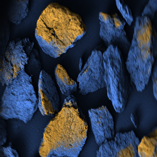
Where the light microscope stops
A light microscope cannot resolve structures finer than about half the wavelength of light. Below that lies the range of the particles we are interested in.
What we do
Every day we breathe air that others have exhaled, along with particles from traffic and industry. Whether – and how much of it – is pathogens or plastic can today only be determined in a laboratory, with considerable effort. We want to make it measurable on site.
About MeiLuftFor researchers
How a scanning electron microscope forms an image, why the sample normally has to go into a vacuum, how our window avoids that – and how our approach relates to established methods, with references.
To the technologyFor investors
Electron microscopy as a service: rent instead of buy, no dedicated laboratory and no specialist staff – for a fraction of what an in-house instrument costs today. One hardware platform, many applications: particles in industry first, without regulatory approval, then ultrafine dust and nanoplastics, and in the medium term pathogens in medicine.
Business model, markets and statusWhat it is needed for
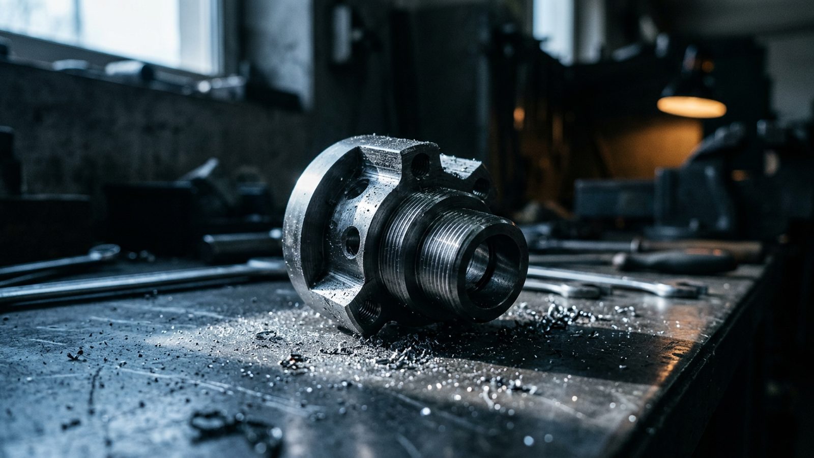
Particles in industry
Seeing abrasion and contamination in manufacturing processes particle by particle, for example in semiconductor and battery production.

Ultrafine dust in the air
Seeing particles individually instead of measuring averages: what they are and where they come from.
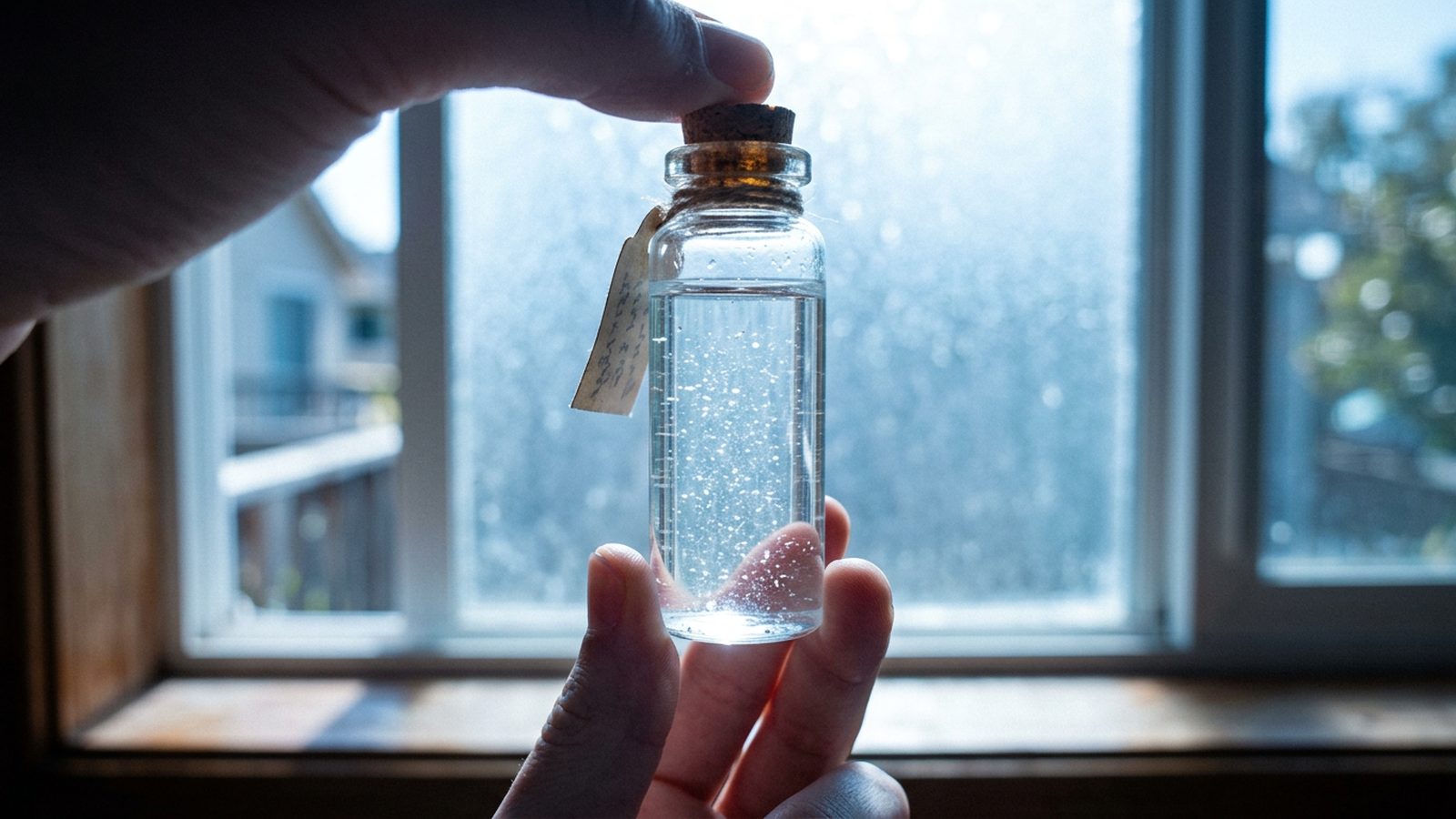
Nanoplastics in water
Imaging plastic particles individually and telling them apart from other particles.
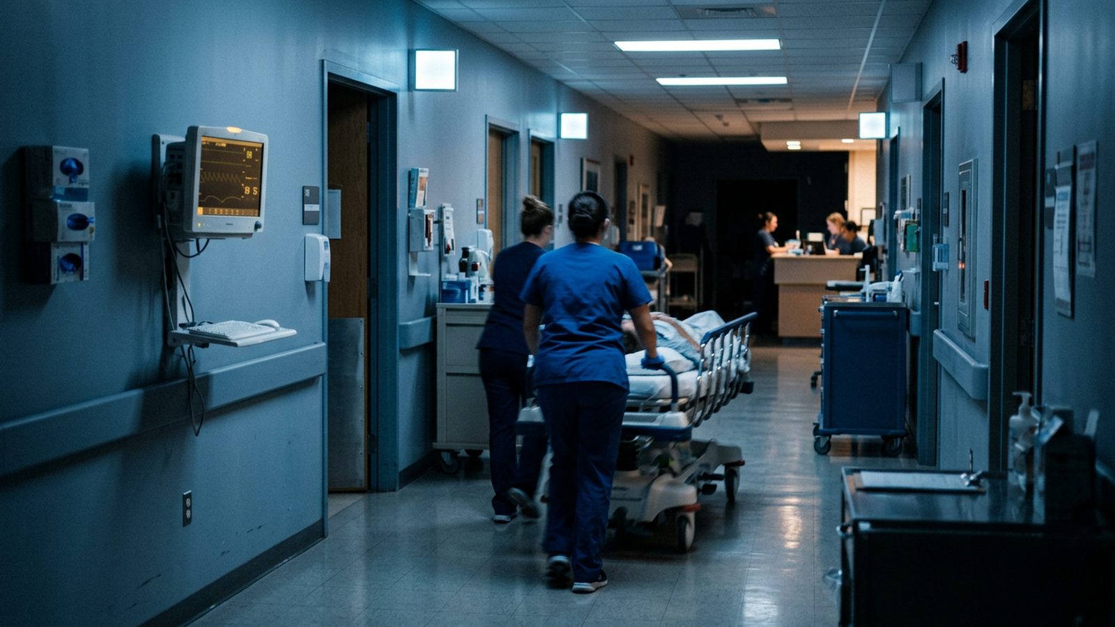
Medicine, in the medium term
Goal: recognising pathogens directly in the sample, for example in sepsis. Subject to regulatory approval, not a current offering.
Images: illustrations, AI-generated.
Where we stand
- done
Electron optics designed
Cross-checked with several independent calculation methods
- done
Beam system tested
In the high-vacuum test rig
- done
First model
Shown at Medica
- done
Physical model
Of the instrument, validated against published measurements
- done
Test microscope
Existing electron microscope with our own electronics and software: first images, automatic particle detection, sample still in a vacuum
- done
Manufacturing documents
For the column of the prototype
- in progress
AI for imaging and detection
Trained on simulated images
- in progress
Prototype
Our own compact instrument with a window: the sample stays in air. Pace depends on funding
- planned next
Measuring in air
Through the window, with the prototype
- planned next
Application studies
And field tests with pilot partners
As of November 2026
Recognition and support
- Research allowance Seal of the German certification body for the research allowance (BSFZ) three years in a row: 2024, 2025 and 2026
- Hohenlohe4Talents Winner 2021, featured in an SWR television report on the final
- Medica Düsseldorf Exhibitor in 2022 and 2023 at the Baden-Württemberg start-up park “The Länd”; in 2023 included in the trade fair’s innovation report
- Partners Baden-Württemberg International, Start-up BW, aformatik